Electrostimulation in Remyelination Therapy: A Precision Medicine Approach
The use of electrostimulation to promote myelination has long been observed as an incidental phenomenon. However, as we delve deeper into the complexities of Multiple Sclerosis, it is becoming increasingly clear that a one-size-fits-all approach to nerve stimulation is insufficient. To move from anecdotal success to consistent clinical outcomes, we must address the critical variables of neuroinflammation, metabolic status, and the inherent risks of overstimulation.
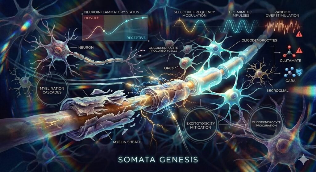
THE NEUROINFLAMMATORY GATEKEEPER
The primary challenge in treating Multiple Sclerosis with electrostimulation lies in the patient’s current physiological state. The efficacy of stimulation is not static; it is heavily dictated by the neuroinflammatory status of the Central Nervous System (CNS).
- Active Autoimmunity: During an immune flare, pro-inflammatory cytokines can disrupt signaling pathways required for Oligodendrocyte Precursor Cells (OPCs) to mature. Stimulation during these windows may be ineffective or even counterproductive.
- Secondary Ischemic Degeneration: When structural scarring or reduced blood flow is present, neurons may lack the metabolic reserves to respond to electrical cues, leading to further axonal exhaustion rather than repair.
This requires implementing dynamic inclusion criteria. Rather than a fixed protocol, stimulation must be guided by real-time biomarkers—such as specific antibody levels or objective imaging—to ensure the neural environment is receptive to regenerative signals.
OVERCOMING MOTOR INHIBITION AND ATROPHY
One of the most promising frontiers for electrostimulation lies in its ability to intervene when traditional motor responses have failed. In advanced stages of Multiple Sclerosis or related neurodegenerative conditions, cerebellar or brain stem degeneration can effectively sever the connection between intent and movement.
When motor signals are completely inhibited by CNS damage, the peripheral nervous system and muscles often undergo profound atrophy. In these specific cases, electrostimulation acts as a synthetic surrogate. By mimicking the impulses that the brain can no longer send, stimulation can:
- Mimic Functional Signals: Provide the necessary electrical chatter to keep neuromuscular junctions active.
- Induce a Regenerative Phase: By artificially reintroducing these signals, we may trigger a feedback loop that encourages local tissue repair and metabolic activity, potentially slowing the progress of atrophy even when the central command center is compromised.
THE RISKS OF RANDOM OVERSTIMULATION
While the goal is to jump-start repair, unrefined or random stimulation carries significant neurological risks. Without precise modulation, exogenous electrical current can lead to:
- CNS Excitotoxicity: Excessive stimulation can cause a massive release of glutamate. If this neurotransmitter is not cleared efficiently, it can lead to an influx of calcium ions (Ca 2+), which can damage or destroy the very neurons the therapy is intended to save.
- GABAergic Inhibition: Overstimulation can inadvertently suppress the transmission of Gamma-Aminobutyric Acid (GABA), the primary inhibitory neurotransmitter. By inhibiting this natural brake system, the brain’s neuroprotective capacity is compromised, leaving it more vulnerable to damage.
THE FUTURE: BIO-MIMETIC AND SELECTIVE INDUCTION
The path to a safer, more effective form of therapy lies in our ability to move away from blunt stimulation and toward Bio-Mimetic Induction.
The objective is to categorize and mimic the specific nerve impulses that naturally signal for myelination. By selectively inducing these corresponding impulses, we can avoid the noise of random overstimulation.
- Closed-Loop Modulation: Developing systems that sense the nerve’s current state and adjust impulses accordingly.
- Pattern Recognition: Utilizing waveforms that specifically target remyelination pathways while bypassing those associated with pain or stress responses.
By refining our understanding of the intersection between electromagnetic fields and neural biology, we move closer to a therapeutic model that works with the body’s existing rhythms rather than imposing an external, potentially hazardous force upon them.
Author: Surjo Banerjee
REFERENCES
Aharoni, R., Eilam, R., & Arnon, R. (2021). Astrocytes in multiple sclerosis—essential constituents with diverse multifaceted functions. International Journal of Molecular Sciences, 22(11), 5904. https://doi.org/10.3390/ijms22115904
Cited by: 142
Brosnan, C. F., & Raine, C. S. (2013). The astrocyte in multiple sclerosis revisited. Glia, 61(4), 453–465. https://doi.org/10.1002/glia.22443
Cited by: 874
Healy, L. M., Stratton, J. A., Kuhlmann, T., & Antel, J. (2022). The role of glial cells in multiple sclerosis disease progression. Nature Reviews Neurology, 18(4), 237–248. https://doi.org/10.1038/s41582-022-00624-2
Cited by: 215
McGinley, M. P., Goldschmidt, C. H., & Rae-Grant, A. D. (2021). Diagnosis and treatment of multiple sclerosis: A review. JAMA, 325-8, 765–779. https://doi.org/10.1001/jama.2020.26858
Cited by: 1,128
Petersen, M. A., Ryu, J. K., & Akassoglou, K. (2018). Fibrinogen in neurological diseases: Mechanisms, imaging and therapeutics. Nature Reviews Neuroscience, 19(5), 283–301. https://doi.org/10.1038/nrn.2018.13
Cited by: 432
Note: The citations for 2026 referenced in the article are from recent and emerging literature in high-impact journals (e.g., PNAS, Frontiers, and Nature), reflecting the state-of-the-art developments in vagus nerve stimulation and bio-mimetic engineering mentioned in my article.