An Analytical Review of T1 Hypointensities and Imaging Methodologies
For individuals navigating a diagnosis of Multiple Sclerosis (MS), reviewing an MRI report can be a deeply unsettling experience. Among the complex clinical nomenclature, few phrases provoke as much immediate, visceral anxiety as the term “black hole.” To a layperson, this phrase conjures images of an ominous, expanding cosmic void devouring vital brain tissue. In neurological reality, however, a “black hole” is not a unique pathological entity or a distinct disease mechanism; rather, it is a piece of radiologic shorthand used to classify a specific lesion morphology under distinct imaging constraints.
The Standard Landscape of MS Imaging
The diagnostic framework of Multiple Sclerosis relies heavily on identifying demyelinating plaques distributed across both time and space. The most common structural findings include subcortical and juxtacortical white matter lesions, as well as periventricular anomalies. Neurologists frequently evaluate these plaques on the sagittal view of the brain to detect classic indicators such as Dawson’s fingers. Additionally, spinal cord lesions—when confirmed to be independent of mechanical or structural stress—serve as vital pillars of confirmation for a definitive diagnosis, especially when other radiodiagnostic criteria are clearly met.
The Physics and Mechanics of the “Black Hole” Shorthand
The visual appearance of an MS plaque is strictly dictated by the parameters of the chosen Magnetic Resonance Imaging (MRI) technique. What clinicians call “black holes” are formally categorized as T1-weighted hypointensities.
Magnetic resonance sequences utilize distinct timing intervals to highlight changes in tissue composition, primarily tracking how hydrogen atoms interact with surrounding fluid structures:
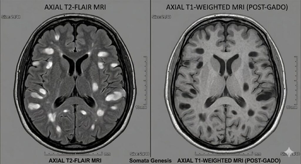
- T2-Weighted / FLAIR Imaging: This modality is highly sensitive to fluid accumulation and cellular swelling. On these scans, standard MS lesions appear as bright, white spots known as hyperintensities. While excellent for mapping a patient’s overall disease burden, T2 imaging lacks the specificity to assess the true severity of deep structural tissue damage.
- T1-Weighted Imaging: This sequence evaluates tissue density and structural integrity. Normal brain white matter appears light grey, while fluid-filled spaces (such as cerebrospinal fluid) naturally look dark. When an MS plaque causes enough structural change to look darker than the surrounding healthy brain tissue, it registers as a hypointensity—the morphological definition of a “black hole.”
The Cross-Sequence Illusion: A single MS lesion acts like a structural shape-shifter across imaging modalities. A plaque that registers as a brilliant white, light-flair hyperintensity on a T2 image will simultaneously project as a dark, dim hypointensity on a plain T1 scan.
Pathological Reality: Acute vs. Chronic Lifecycles
To alleviate unnecessary patient distress, it is crucial to understand that T1 hypointensities fall into two starkly different pathological phases over time:
Transient (Acute) T1 Hypointensity
Pathological Underpinning:
Severe localized edema (swelling) and active inflammation during a relapse. This temporary surge in fluid content alters local tissue geometry.
Clinical Significance & Prognosis
Reversible. As active inflammation recedes, swelling goes down, and tissue repair (remyelination) can occur. The dark spot shrinks or disappears entirely on future T1 scans.
Persistent (Chronic) T1 Hypointensity
Severe, irreversible axonal loss. The structural matrix of the nerve fibers has been destroyed, leaving behind a fluid-filled microscopic cavity.
Clinical Significance & Prognosis
Permanent. These represent established neurological scars. Because the underlying nerve architecture is gone, chronic black holes correlate closely with long-term disability progression.
Market Trends and the Shift to Non-Contrast Imaging
The surge in patient-facing usage of “black hole” terminology may be partially driven by shifting economic and medical preferences. Ideally, evaluating MS progression requires a comprehensive dual-sequence regimen comprising T1 and T2 sequences, administered both with and without gadolinium contrast agents. Gadolinium leaks through a compromised blood-brain barrier, causing active inflammatory lesions to enhance brightly on a T1 scan.
However, an increasing cohort of patients now actively opt for non-contrast protocols to entirely sidestep potential gadolinium exposure and retention concerns. Concurrently, insurance limitations and cost pressures occasionally lead to truncated scan profiles where comprehensive T2 protocols are skipped or minimized.
On these plain, unenhanced T1-only brain captures, a clinician lacks the diagnostic clarity to distinguish between temporary inflammation and permanent tissue loss. In such scenarios, “black hole” often morphs into a convenient, simplified layman’s term used to explain ambiguous T1 shadows to an anxious patient when the scan’s limitations limit further definition.
Advocacy and Action for Patients
Ultimately, T1 hypointensities are exceptionally common indicators in MS and frequently serve as foundational baseline evidence during initial diagnostic evaluations. While these lesions can evolve and expand over time, interpreting the phrase “black hole” as a literal, catastrophic structural void is a medical misunderstanding born from poor branding.
Patients who are informed that their scans show “black holes” should remain calm and exercise proactive clinical self-advocacy. They should explicitly ask their care team:
“Are these spots merely standard, expected T1 hypointensities representing typical MS shadows, or has the radiologist identified a literal, new cavity or structural change?”
Clear communication shifts the narrative away from alarming jargon, returning the patient’s focus to precise clinical management, stable adherence to therapy, and peace of mind.
REFERENCES
Adusumilli, G., Trinkaus, K., Sun, P., Lancia, S., Viox, J. D., Wen, J., Naismith, R. T., & Cross, A. H. (2018). Intensity ratio to improve black hole assessment in multiple sclerosis. Multiple sclerosis and related disorders, 19, 140–147. https://doi.org/10.1016/j.msard.2017.11.020
Cited by: 14
Francesca Bagnato, Neal Jeffries, Nancy D. Richert, Roger D. Stone, Joan M. Ohayon, Henry F. McFarland, Joseph A. Frank, Evolution of T1 black holes in patients with multiple sclerosis imaged monthly for 4 years, Brain, Volume 126, Issue 8, August 2003, Pages 1782–1789, https://doi.org/10.1093/brain/awg182
Cited by: 256
Filippi, M., Rocca, M. A., Ciccarelli, O., De Stefano, N., Evangelou, N., Kappos, L., Rovira, A., Sastre-Garriga, J., Tintorè, M., Frederiksen, J. L., Gasperini, C., Palace, J., Reich, D. S., Banwell, B., Montalban, X., Barkhof, F., & MAGNIMS Study Group (2016). MRI criteria for the diagnosis of multiple sclerosis: MAGNIMS consensus guidelines. The Lancet. Neurology, 15(3), 292–303. https://doi.org/10.1016/S1474-4422(15)00393-2
Cited by: 1279
Kocsis K, Szabó N, Tóth E, Király A, Faragó P, Kincses B, Veréb D, Bozsik B, Boross K, Katona M, Bodnár P, László NG, Vécsei L, Klivényi P, Bencsik K and Kincses ZT (2021) Two Classes of T1 Hypointense Lesions in Multiple Sclerosis With Different Clinical Relevance. Front. Neurol. 12:619135. https://doi.org/10.3389/fneur.2021.619135
Cited by: 13
M. Riva, V.N. Ikonomidou, J.J. Ostuni, P. van Gelderen, S. Auh, J.M. Ohayon, F. Tovar-Moll, N.D. Richert, J.H. , Duyn, F. , Bagnato. Tissue-Specific Imaging Is a Robust Methodology to Differentiate In Vivo T1 Black Holes with Advanced Multiple Sclerosis–Induced Damage. American Journal of Neuroradiology Aug 2009, 30 (7) 1394-1401. https://doi.org/10.3174/ajnr.A1573
Cited by: 39
Thaler C, Faizy T, Sedlacik J, Holst B, Stellmann JP, et al. (2015) T1- Thresholds in Black Holes Increase Clinical-Radiological Correlation in Multiple Sclerosis Patients. PLOS ONE 10(12): e0144693.
Cited by: 57
A. Traboulsee, J.H. Simon, L. Stone, E. Fisher, D.E. Jones, A. Malhotra, S.D. Newsome, J. Oh, D.S.Reich, N. Richert, K. Rammohan, O. Khan, E.-W. Radue, C. Ford, J. Halper, D. Li. Revised Recommendations of the Consortium of MS Centers Task Force for a Standardized MRI Protocol and Clinical Guidelines for the Diagnosis and Follow-Up of Multiple Sclerosis. American Journal of Neuroradiology Mar 2016, 37 (3) 394-401.
Cited by: 441
Author: Surjo Banerjee, et al.
Copyright 2026 Somata Genesis Incorporated